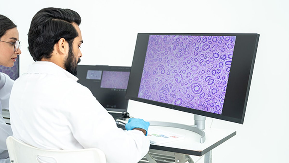
During the COVID-19 pandemic, the need for remote work pushed digital pathology from a niche tool to a core component of laboratory workflows. By scanning glass slides into high-resolution images, pathologists could continue diagnoses without physically accessing microscopes, and the resulting files could be shared instantly across continents, enabling real-time collaboration among experts who were otherwise isolated.
Digitized slides become data assets
After the initial shift, researchers began extracting additional information from the pixel data. “Before, the pathology endpoint was just a report,” said Aleksandra Zuraw, DVM, PhD, a veterinary pathologist at Charles River Laboratories. “Now you have the digitized slide, which is pixels.” She emphasized that each image carries layers of visual detail that were previously inaccessible without manual re-examination.
The same archived tissue can be revisited for molecular assays. When a fresh hypothesis emerges after a study ends, scientists can run tests on the stored formalin-fixed, paraffin-embedded (FFPE) blocks instead of launching a new animal experiment, thereby preserving valuable resources and respecting ethical considerations.
In April 2024, Charles River Laboratories launched its Alternative Methods Advancement Project, aiming to extract more value from each animal used in preclinical studies. One short-term goal is to replace some live control animals with “virtual control groups” built from matched historical data, a strategy that relies on robust statistical matching and careful documentation of prior study conditions.
Regulatory push and new technologies
FFPE blocks preserve not only histology but also molecular signatures of drug response. While fresh-frozen tissue is ideal for omics work, it must be collected prospectively. The Organisation for Economic Co-operation and Development’s 2025 guidance highlights that properly stored FFPE material can support gene-expression and protein studies, provided preparation and storage are consistent across sites.
Read Also: New Drug Survodutide Shows Promise for Obesity, Diabetes
The U.S. Food and Drug Administration’s 2022 Modernization Act 2.0 allowed nonanimal methods to support applications to begin human drug trials. In April 2025, the agency published a roadmap for reducing animal testing in preclinical safety studies, starting with monoclonal antibodies and later expanding to other biologics and eventually new chemical entities. Over three to five years, the agency aims to make animal studies the exception rather than the norm, with new approach methodologies (NAMs) as the default.
Parallel advances include molecular prediction from standard hematoxylin and eosin (H&E) stained slides. Researchers can apply emerging algorithms directly to digitized H&E images, extracting information without additional laboratory steps. Virtual staining, which generates a stained appearance from an unstained scan, is also gaining traction and may eventually eliminate the need for glass slides altogether, simplifying logistics and reducing waste.
NYU Pathology Department Shifts Entirely to Digital Sign-Out
In a recent podcast, Zuraw interviewed Syed T. Hoda, M.D., NYU’s director of digital pathology, about moving roughly 95 pathologists to digital sign-out in a single rollout.
Zuraw noted that virtual staining is expanding quickly, potentially letting labs skip both staining and glass slides. A 2026 review documents an accelerating proliferation of virtual-staining applications in research and clinical settings across a range of tissue types, tracing back to 2019 demonstrations.