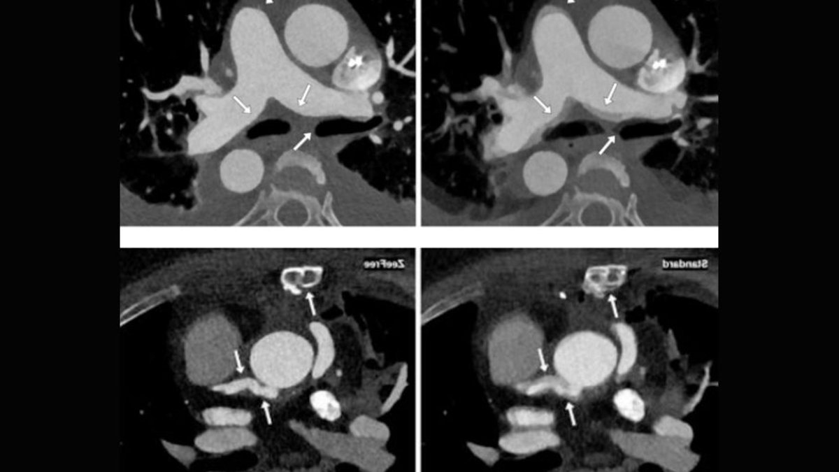
A new motion correction technique demonstrates significant potential to minimize breathing-induced distortions in computed tomography angiography (CTA) scans performed shortly after coronary artery bypass graft (CABG) procedures. The method enhances visualization across bypass grafts, native coronary vessels, and additional thoracic anatomy.
The innovation tackles a common issue in early postoperative imaging, where patients often face challenges sustaining a steady breath-hold during scanning. Additionally, evaluating bypass grafts necessitates broader anatomical coverage than typical cardiac CT protocols. Among patients whose initial scans exhibited notable respiratory motion artefacts, the technique successfully transformed all previously unassessable anatomical areas into diagnostically useful images.
Restoring Clarity to Compromised Scans
When two independent reviewers assessed 58 regions initially deemed unusable due to breathing motion, every single area became evaluable after applying the correction algorithm. Most of these regions achieved the highest possible image quality rating.
The improvements extended well beyond the bypass grafts alone. Significant reductions in artefact severity were observed across all examined structures, including native coronary arteries, the aorta, pulmonary trunk, lungs, trachea, and sternum. Previously distorted images characterized by double contours, blurring, and discontinuities showed markedly improved anatomical consistency following correction.
Challenges in Early Postoperative Imaging
Respiratory movement poses a substantial technical hurdle when performing CTA shortly after CABG surgery. Recovering patients frequently struggle to maintain the prolonged breath-hold required for optimal image acquisition, while full bypass graft assessment demands wider anatomical coverage than standard cardiac CT examinations.
Read Also: Global Survey Finds CT Patient Centering Inconsistent
In a study of 680 consecutive patients undergoing early postoperative CTA, 80 individuals exhibited significant breathing artefacts on standard reconstructions. The correction technique was applied exclusively to this subgroup where respiratory motion was clearly visible. Five patients lacked necessary raw data for retrospective processing, leaving 75 patients with 205 bypass grafts available for analysis. These scans occurred at a median of four days following surgery.
The algorithm functions by identifying and correcting misalignments between adjacent image slices through overlapping acquisition data. It calculates spatial displacements and realigns affected image sequences to create a more coherent volumetric representation. Originally developed to address stair-step artefacts caused by heart rate variability, the same methodology proved effective in reducing respiratory motion-induced discontinuities.
Unanswered Clinical Questions Remain
While the technical image quality enhancement suggests potential utility for motion correction in early postoperative CTA, its actual clinical value remains undefined. The current evaluation focused solely on artefact reduction and image quality metrics rather than assessing graft stenosis detection or occlusion identification.
The findings originate from a single academic medical center and involve a limited patient cohort. The study utilized only one scanner model and a proprietary correction algorithm. Whether comparable benefits would emerge with different imaging systems, acquisition protocols, or in varied clinical environments remains unverified.
Motion correction technology has shown promising results in enhancing CTA image quality when respiratory artefacts affect patients undergoing early postoperative evaluation after CABG surgery. The approach successfully reduced artefacts across both cardiovascular and non-cardiovascular thoracic structures, restoring diagnostic capability in previously unassessable regions. However, current evidence demonstrates technical improvement in a specific patient group rather than proven clinical benefits.