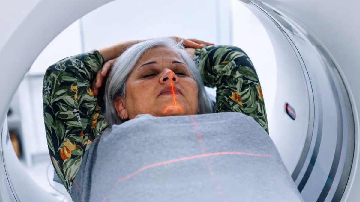
A global study involving 321 radiographers from 43 nations revealed significant inconsistencies in CT patient centring practices worldwide. Participants described frequent positioning mistakes, varying expertise regarding radiation dose implications, and restricted adoption of automated alignment systems. The diverse sample, covering hospitals, imaging facilities, and academic centers, offered broad insights into real-world scanning procedures.
Vertical misalignment below the CT gantry’s central axis emerged as the most common error type. Respondents also showed differing levels of understanding about how localiser image orientation and off-centre positioning influence radiation exposure and Hounsfield unit measurements. These results show discrepancies between theoretical instruction and the challenges faced during actual scanning operations.
Positioning Inconsistencies Continue
Precise patient alignment with the CT gantry’s central axis is critical because automatic tube current adjustments depend on accurate size and attenuation assessments. Any off-centre placement can interfere with dose modulation, degrading image quality or increasing radiation levels unnecessarily.
Survey participants most frequently cited vertical positioning errors below the isocentre as the primary issue, though lateral and above-axis misalignment were also noted. Follow-up scans compounded the problem, with nearly 40% of radiographers estimating that fewer than 10% of returning patients were positioned identically across visits. This variability suggests even experienced technicians may struggle to maintain consistent placement between examinations.
Read Also: Pharmaceutical convention to focus on AI and digitalisation
Knowledge Deficits Impact Dose and Image Measurements
Awareness of mis-centring’s consequences varied widely among respondents. While most recognized automatic tube current modulation as essential for dose control and understood its dependence on patient attenuation, fewer grasped how positioning affects the system’s dose-adjustment algorithms.
Automated Centring Tools See Limited Adoption
While some clinical settings have access to automated centring technology, its use remains sporadic. Only about one-third of survey participants reported having such systems available, with 3D camera-based solutions being the most prevalent option. Facilities without automation typically relied on manual visual cues and anatomical landmarks.
Respondents viewed automation positively for its potential to enhance image quality, reduce radiation exposure, improve workflow efficiency, and ensure scan reproducibility. The most highly rated benefit was lower radiation doses. However, practical obstacles, including operational complexity and over-reliance on technology, often discouraged widespread implementation.
The survey frames automation as a supplementary aid rather than a complete replacement for professional positioning expertise. Some respondents warned that over-reliance on automated systems could diminish careful attention to patient placement, potentially reintroducing the very errors these tools aim to prevent.