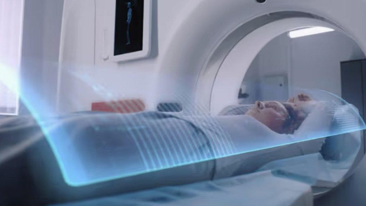
Adjusting tube voltage downward presents an effective strategy for minimizing radiation during contrast-enhanced abdominal and pelvic CT scans, all while maintaining the clarity needed for accurate diagnosis. Data from 13 studies demonstrates that protocols set at 80 kVp, when combined with noise-reduction reconstruction methods, can achieve meaningful dose savings.
Automated voltage selection and size-based protocols can further tailor exposure levels. Still, results vary depending on the scanner model, reconstruction approach, and individual patient factors. Current research has yet to confirm whether improved image appearance at lower voltages consistently translates into better detection of subtle or faint lesions.
Advanced Reconstruction Expands Low-kVp Use
The primary hurdle when lowering tube voltage is managing increased image noise. While reduced kVp levels cut radiation and may improve contrast by boosting iodine visibility, the weaker X-ray energy can degrade image sharpness if not properly compensated. Modern reconstruction tools have helped address this trade-off. Hybrid iterative reconstruction has become standard for noise suppression in low-kVp scans, while model-based iterative methods enable further dose reductions without sacrificing diagnostic utility. Recently, deep learning reconstruction has entered the picture, offering enhanced noise reduction and finer spatial detail to support clearer imaging at lower doses.
Personalised Protocols Could Improve Dose Optimisation
One-size-fits-all low-kVp approaches are impractical. Patient body size directly influences noise levels, dictating how much voltage reduction remains viable while keeping images diagnostically useful. Body mass index (BMI) remains the most common factor in protocol selection.
Read Also: New Technique Clears Breathing Artifacts in Post-CABG Scans
Studies using BMI-based adjustments have successfully lowered radiation while preserving image quality across diverse patient groups. Manually adjusting voltage by size has also proven effective, though automated systems provide a more consistent way to adapt exposure based on tissue density and contrast levels. Pairing voltage selection with automatic current modulation adds another layer of personalization. However, kVp alone does not dictate total radiation, tube current and the number of scan phases also play significant roles, meaning dose optimization requires a broader approach.
Image Quality Does Not Guarantee Lesion Detection
Yet visually sharp images do not automatically ensure equal diagnostic effectiveness. Research on heavily reduced-dose scans shows that deep learning can enhance perceived quality and contrast ratios, even as it may impair the visibility and assessment of small, low-contrast liver lesions.
To refine low-kVp strategies for wider adoption, the field requires standardized reporting, performance evaluations tied to specific clinical tasks, and large-scale multicenter trials.